This here is a presentation with associated notes that we put together on the subject of mechanical ventilation stratgies for different types of congenital heart conditions. We made it based on thoughts and data we could glean from literature (see refs at the very end) and what we already knew about mechanical ventilation, but it has not yet been reviwed by any sort of specialist or subject matter expert. We’re putting it up here to both increase visibility so that hopefully we can get such a review, but also because we mention it very briefly in the next version of the vent book and didn’t actually have it up on the site anywehere for curious minds. In any case, it’s good food for thought and a decent review of different conditions.
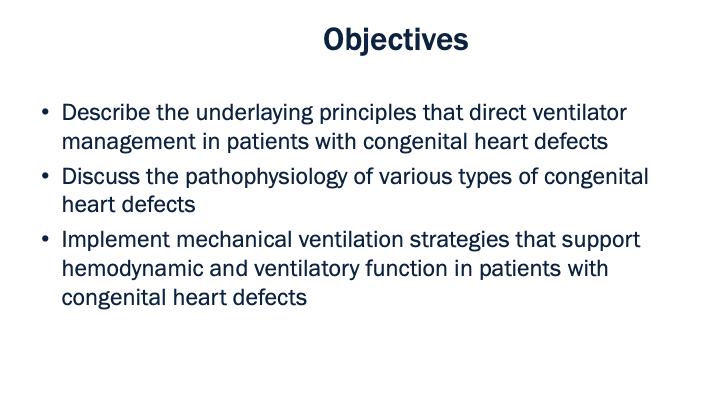
Before we get into the specifics of ventilator management, there are a few things to review. We’ll discuss each of the items listed here, beginning at the top of the list.
So to start with: what is a congenital heart defect or CHD? In simple terms it’s when the heart, and therefore the connected circulatory components, are functionally different than normal at birth. There are many, many different types of congenital heart defects, but we tend to organize them by groups of conditions that share similar pathology. For example, we’ll talk later about defects that lend themselves to either increased or decreased pulmonary blood flow and we’ll mention the idea of “mixing lesions” – these are simplifications that help us identify treatment strategies when time is of the essence.
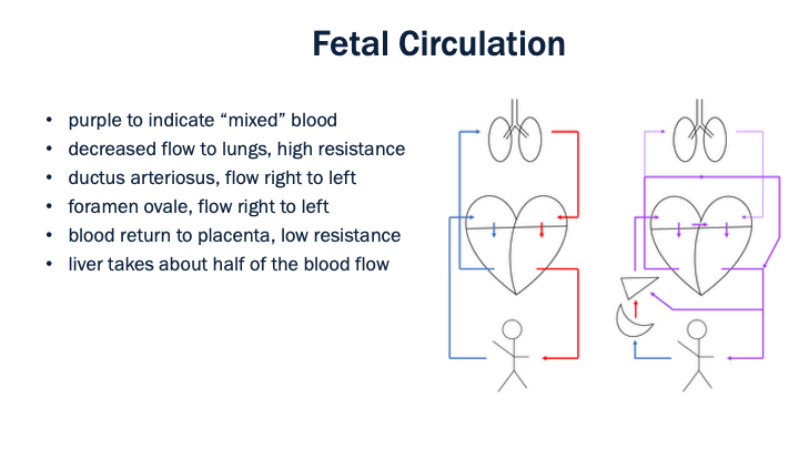
The next Underlaying Principle to discuss is the process by which circulation transitions from fetal to neonatal blood flow. This should be a review, but we’ll outline a few details just to make sure we are all on the same page. The first graphic, with all of the arrows in either blue or red, represents normal circulation. The one on the right, with purple arrows and a few extra items, represents fetal circulation in utero.
Important differences to note between the two are listed on the slide. First is the fact that blood moves through the fetus in a ”mixed” or not-fully-oxygenated state. The crescent shape on the bottom left of the fetal circulation graphic is the placenta – that’s where oxygen gets onloaded into fetal circulation. Roughly half of that oxygenated blood passes through the liver and then what gets to the heart is in this “mixed” state. The details of that portal circulation system aren’t particularly relevant for this discussion, so enough said there. It’s also worth noting that not all of this purple or “mixed” blood is equally oxygenated, this is a simplification of a complex process.
Next idea: blood leaving the right ventricle in the normal human (pictured left) goes to the lungs. In fetal circulation, however and on the right, there is a high degree of resistance to blood flow in the lungs as well as a bridge between the pulmonary artery and the aorta (the ductus arteriosus). The result is that blood flows directly from the pulmonary artery to the aorta and bypasses the lungs more or less altogether. The graphic shows this as a roundabout path, but in reality these tow vessels pass quite close to one another and the ductus arteriosus is quite small in length.
There is another alternative pathway for blood in the fetal heart between the right and left atria called the foramen ovale. In both the foramen ovale and the ductus arteriosus, blood flow in normal fetal circulation is from the right side of the heart to the left side of the heart (as depicted by the arrows in the graphic on the right).
And then very last thing to note is that in normal fetal circulation resistance to blood flow at the placenta is very low. This allows blood to return back to the placenta from systemic circulation and thus repeat the cycle all over again.
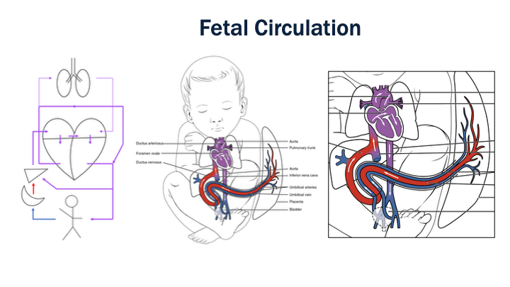
Before moving on, we’ll just point out one quick thing here. We mentioned on the last slide that we’ve drawn, in our simplified sketch, the ductus arteriosus as a long route from the pulmonary vein over and around to the aorta. In reality, however, nature is much more elegant. You can see in these graphics on the right that the pulmonary artery and aorta sort of intertwine with one another and that the bridge formed by the ductus arteriosus is actually very compact.
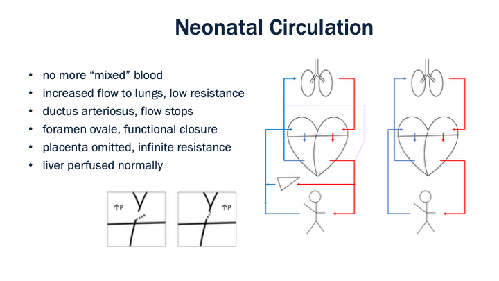
Now once the fetus leaves the womb and becomes a neonate things change a bit. Within the first few minutes of life circulation normally begins to resemble what we’d see in an adult patient. That said, the ductus arteriosus and foramen ovale are still present, as indicated by the dotted purple lines.
First of the points to mention regarding neonatal circulation: no more “mixed” blood. We previously saw the oxygenation step happening at the placenta during fetal circulation, but that ceases to exist along with hepatic blood flow via a mechanism we said we weren’t going to delve too far into. So we have oxygenation occurring in the lungs just like we’d see in an adult. To describe this a bit more technically: blood flow increases to the lungs as resistance to that blood flow decreases.
Next thing to point out is that both the ductus arteriosus and the foramen ovale eventually close. The ductus arteriosus normally closes within a day. The foramen ovale takes much longer to close, on the order of months, but it actually exists as a sort of flap opening into the left atrium, so as pressure in that chamber increases with the transition to normal circulation, it essentially shuts the door to any left-to-right shunt.
So this is how it all happens normally. One more Underlaying Principle and then we’ll talk about what can go wrong.
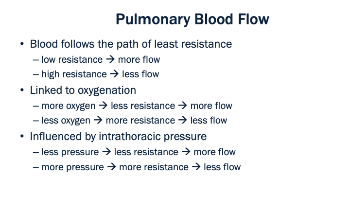
Understanding pulmonary blood flow in the neonate is absolutely crucial to implementing ventilation strategies in the presence of congenital heart defects, so let’s take a moment to work through this concept. Starting with the basics, blood will flow to places with decreased resistance. Resistance in terms of blood flow implies changes in vasculature – vasodilation decreases resistance and essentially invites more blood flow, vasoconstriction increases resistance and pushes blood away to other regions. Easy enough, so lets move on.
In the lungs we have this effect called hypoxic pulmonary vasoconstriction. Hypoxic pulmonary vasoconstriction describes the idea that decreased levels of oxygen in the lungs leads to vasoconstriction. This is the exact opposite of what happens in systemic circulation where hypoxemia causes vasodilation. Areas of the lung with less oxygen experience vasoconstriction (i.e. more resistance) and therefore less blood flow. This mechanism helps the body to avoid wasting precious resources in pulmonary circulation: if blood flow were increased to area of low oxygenation, it would pass through without taking oxygen on and the result would be what we call a shunt (which is a type of V/Q mismatch). So less oxygen equals less flow via this phenomenon we call hypoxic pulmonary vasoconstriction.
And then lastly we know that pressure in the chest cavity also effects blood flow. More pressure in that area means less blood flow to the lungs. In terms of mechanical ventilation we often attribute this effect to positive end-expiratory pressure (PEEP), but it can also be considered in terms of mean airway pressure (MAP). So more of either one leads to less flow at the lungs.
note: pulm VR decreases over 6-8 weeks (which increases shunt –> HF)
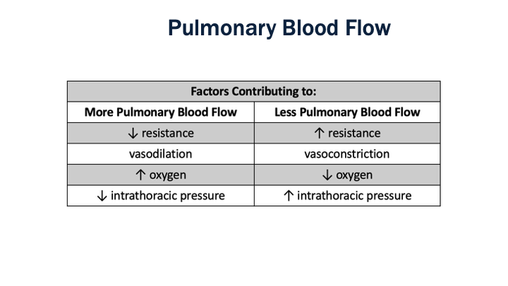
This slide here is just a summary of what we discussed in the last slide in a different format. As we said before, this is very important to understanding how we manage different defects, so if this still isn’t clear please go back and review the last bit. At the very least consider committing this chart to memory by writing it down or taking a screenshot - having it to refer to will help work through some of the ideas in the next section.

There are over thirty different types of congenital heart defects, but we often simplify that and organize them into a few groups as seen on this chart. It is worth noting, however, that these categorizations are simplifications and that any of these defects can occur on a spectrum of severity – our role in management is to maintain physiologic function and some of these defects will ultimately require surgical correction.
And then it is worth noting that we start our discussion of ventilation strategy for patient with CHD at those first two distinct rows and then adjust as needed based on patient presentation. The result is an initial strategy based on type of shunt in the context of pulmonary blood flow, then we adjust based on other factors. This will all become clear soon enough.
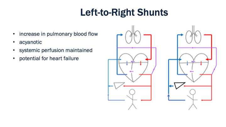
The first collection of defects to discuss are those that cause an increase in pulmonary blood flow. As shown by each of the three purple lines, this could refer to a patent ductus arteriosus (PDA), an atrial septal defect (ASD) or a ventral septic defect (VSD). In any one of these cases blood is shunted from the left side of the heart to the right side. The result is that oxygenated blood coming from the lungs is redirected back into pulmonary circulation. Oxygenation is fine and the patient presents without marked cyanosis, but perfusion can be negatively affected if the shunt is significant
One potential problem in all of this is that the redirection of blood back into pulmonary circulation can eventually lead to pulmonary hypertension and/or heart failure if the heart can’t keep up with the increased workload. The most extreme manifestation would be if pressures on the pulmonary side exceed pressures on the system side and blood flow reverses through the shunt. This phenomenon is called Eisenmenger syndrome – it is quite rare and would present with acute hypoxia with cyanosis.
The timeline at which the progression from increased pulmonary blood flow to heart failure occurs depends on which specific defect is present, the severity or degree of the defect, and the age of the child. Clinical presentation may not occur for weeks, months or years after birth. In most cases, however, these shunts close with time and there is no permanent or lasting consequence.
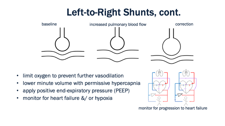
Management of the patient with a left-to-right shunt is focused on avoiding further shunting of blood or exacerbation of the pathology. Oxygen can cause further vasodilation and increase the shunt effect, so should be avoided unless there is evidence of hypoxia. Ventilation should be titrated down to maintain an EtCO2 slightly higher than normal – this will both prevent further vasodilation and maintain cerebral oxygen delivery. And then we can also utilize PEEP to increase intrathoracic pressure and prevent the shunt from worsening.
An important distinction in all of this is the difference between increased pulmonary blood flow and pulmonary hypertension. These are not the same and it’s worth clarifying. More blood into the pulmonary vasculature paired with vasodilation of the vasculature won’t necessarily cause pulmonary hypertension. On the other hand, increased pulmonary blood flow paired with vasoconstriction or inadequate left heart function can lead to increased pressure and eventually heart failure. So while we address the left-to-right shunt by making changes to mitigate the shunt, we must continually monitor for evidence of heart failure and tailor interventions if needed. While the timeline over which this could occur likely excludes it from happening in transport, these problems may occur prior to our patient contact.
All that said, if there is evidence of heart failure, acute pulmonary hypertension, or hypoxia treatment would shift to address those causes. We’ll come back to the management of those complications in just a bit.
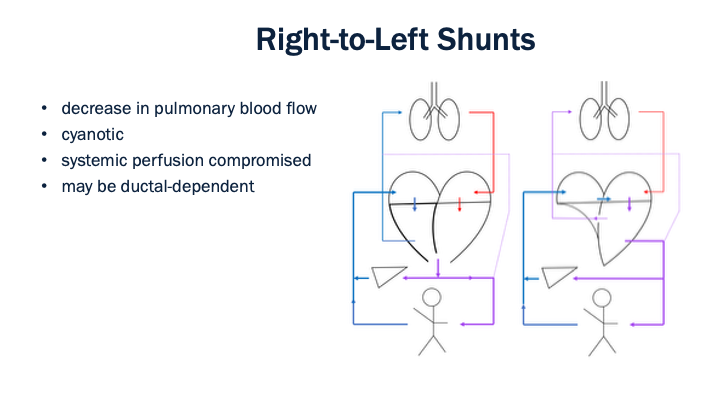
The next situation to discuss is when there is a decrease in pulmonary blood flow and a right-to-left shunt. The two primary examples of this are Tetralogy of Fallot (on the left) and tricuspid atresia (on the right). There is some variation in the presentation of each of these conditions, but just to review the pathophysiology:
In Tetralogy of Fallot is the combination of four findings: pulmonary stenosis, right ventricular hypertrophy, ventricular septal defect and an aorta that leaves the heart from an origin points at that ventricular septal defect.
In tricuspid atresia, on the other hand, the tricuspid valve does not develop appropriately, there for blood goes from the right atrium to the left atrium via an atrial septal defect. In most cases there is some blood flow from the left ventricle through a ventricular septal defect into the right ventricle, but that right ventricular is hypoplastic and smaller than normal.
In either case the result is a decrease in pulmonary blood flow that results in inadequate oxygenation and hypoxia. These are often referred to as cyanotic defects and each case, depending on severity, can be ductal-dependent as well. In cases where oxygenation is dependent on blood flow from to the lungs via the ductus arteriosus, acute deterioration will typically happen shortly after birth and would require prostaglandin infusion from a sending facility prior to transport.
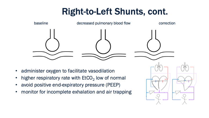
Treatment for these conditions, in terms of mechanical ventilation, is focused on improving oxygen delivery. That said, the mechanism by which we go about that is a bit different than with the typical hypoxic patient. We can still use FiO2 and give oxygen up to 100% as needed – this will cause vasodilation of the pulmonary vascular bed and increase blood flow to the lungs. As for PEEP, however, we want to avoid increasing intrathoracic pressures and should limit PEEP to a physiologic normal or aim even lower than that. Another consideration is to increase ventilation to decrease EtCO2 - this will both facilitate a further decrease in pulmonary vascular resistance and improve oxygen delivery. We make this happen by increasing respiratory rate.
And then following along with this idea, we need to ensure that complete exhalation occurs with an increase in respiratory rate. Monitor for air trapping or AutoPEEP and consider extending the expiratory time (by decreasing the inspiratory time or directly manipulating the I:E ratio) if needed.
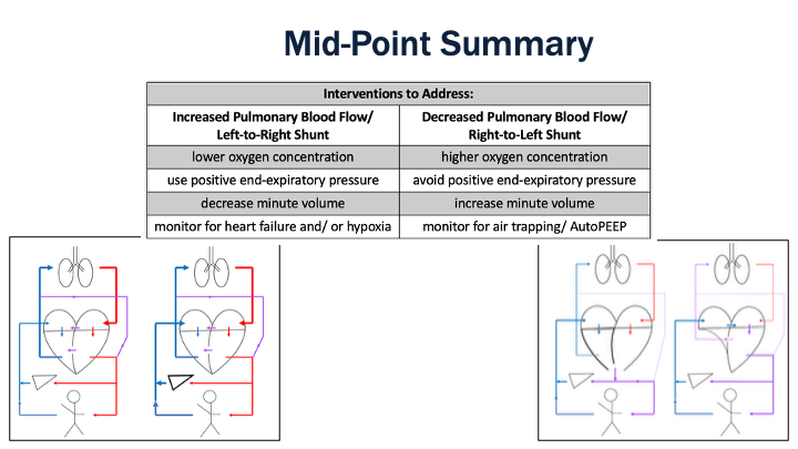
Just to summarize up to this point, there are two problems we have discussed so far.
On the left we have things like patent ductus arteriosus (PDA), atrial septal defect (ASD), and ventricular septal defect (VSD). The issue in these situations is a left-to-right shunt characterized by increased pulmonary blood flow and low pulmonary vascular resistance. Interventions are focused on increasing that pulmonary vascular resistance and we must be vigilant for evidence of heart failure or acute hypoxia. This is our baseline strategy for acyanotic heart defects.
On the right we have Tetralogy of Fallot and tricuspid atresia, both of which result in a right-to-left shunt and are characterized by decreased pulmonary blood flow and higher pulmonary vasculature resistance. Interventions here are focused on decreasing pulmonary vascular resistance and improving oxygenation. That said, we need to avoid causing an unnecessary increase in intrathoracic pressure by both limiting PEEP and monitoring for air trapping. This is our baseline strategy for cyanotic heart defects.

Looking back at our chart from earlier, we’ve covered the first two rows and that gives us our two baseline strategies for ventilator management with congenital heart defects. The next row summarizes obstructive lesions, which is the next area of focus. Looking at the list on the far right of the chart we see that some of those are on the left side of the heart (aortic stenosis and coarctation of the aorta) and the other is on the right side (pulmonary stenosis). Regardless of specific pathology, however, the main concern is a low perfusion state, so maintaining perfusion and preventing a shock state is the focus of treatment for obstructive lesions.
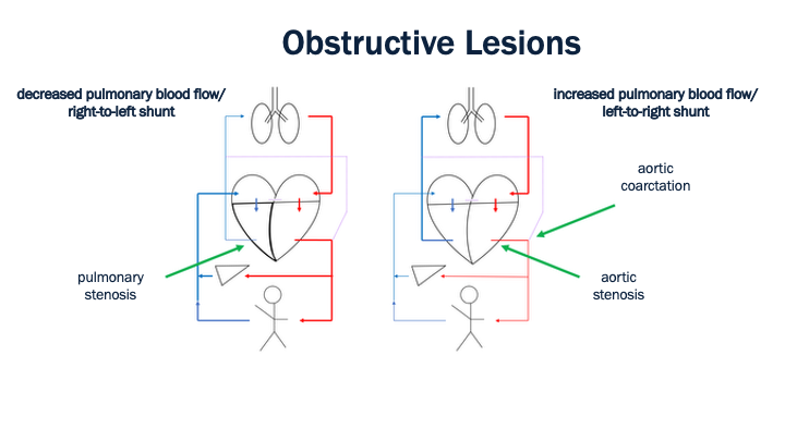
That said, the pathophysiology in each shock state is a bit different. In the case of pulmonary stenosis the problem is decreased blood flow to the lungs and, therefore, an issue with onloading of oxygen that is best treated as we did with other states of decreased pulmonary blood flow: oxygen, minimal PEEP, minute volume above normal with and goal EtCO2 low of normal.
In the case of the case of either aortic stenosis or aortic coarctation the shock issue is with getting enough blood flow past the physical obstruction to perfuse the system effectively. Ultimately this can lead to a left-to-right shunt and we treat it as we did for other cases of increased pulmonary blood flow: limit oxygen if possible (but know that judicious use to address severe hypoxia is OK), utilize PEEP (especially with pulmonary edema), decrease minute volume and aim for an EtCO2 a bit higher than normal.
It is also worth noting that all of these obstructive lesions occur on a spectrum of severity and may be associated with other pathologies. The list of possibilities makes it a bit difficult to identify more nuanced vent strategies, so instead we focus on identifying whether there is evidence of a shunt as previously discussed and then tailoring ventilator management as needed (which we will discuss shortly).
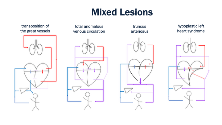
The last category of congenital heart defects to discuss are the mixed lesions or mixing lesions. Examples of these are shown above with graphic representations of common structural anomalies, but the details in each case vary by severity. As a starting point, we can use the decreased pulmonary blood flow and increase pulmonary blood flow frameworks to manage these patients, just as with the obstructive lesions. The difference here is that these mixed lesions tend to present with decreased pulmonary blood flow and, therefore, cyanosis.
The difference, however, is that we must be a bit more judicious in the application of oxygen, as these mixed lesions tend to be ductal-dependent and oxygen can promote closure of the ductus arteriosus. That said, the timeline at which that occurs is not immediate, so it is OK to administer oxygen to address severe hypoxia, we just need to keep in mind that a lower SpO2 goal is completely appropriate.
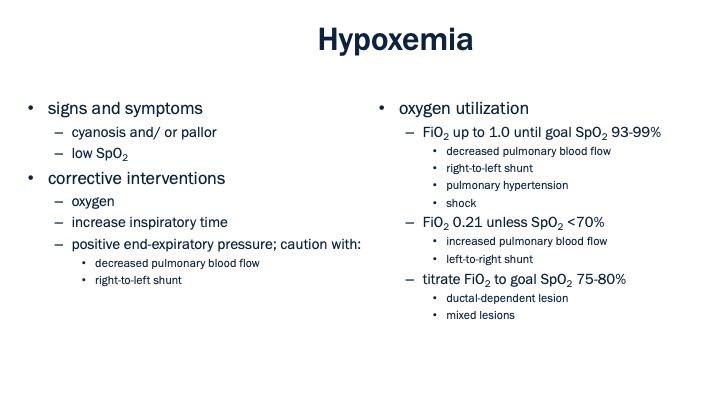
To expand on this idea, let’s move beyond the discussion of specific conditions and consider general guidance for addressing hypoxemia in these patients. We’ve already mentioned that one difference in our normal treatment approach is the use of positive end-expiratory pressure or PEEP – PEEP can improve oxygenation but may have negative consequences.
Another difference, to sort of summarize things we’ve touched on along the way, is how, exactly, to use oxygen or titrate the fraction of inspired oxygen (FiO2) for these patients. Specific guidance is shown there on the right. This should all be a review at the macro level, but specific ranges are provided here.
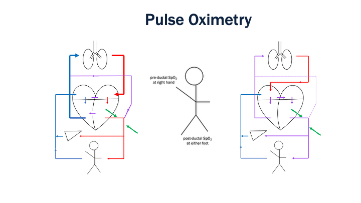
As a point of clarification on the use of pulse oximetry in neonates and/ or infants, standard practice is to measure an SpO2 in two locations – at the right hand or pre-ductally and at either foot or post-ductally. As you can see on the left, if there is shunting of blood from left-to-right via a patent ductus arteriosus, blood goes away from that junction in between the pre and post-ductal reading, so no mixing occurs and the values will be similar. On the right where we have a right-to-left shunt, blood can move into or towards that junction (between the green arrows, indicating blood flow to each SpO2 site) via the PDA – this results in mixing and a potentially lower reading afterwards or post-ductally. This is a generalization, however, and the reality of the situation here depends a lot on the severity of shunt. It’s also worth noting that a difference in pre and post ductal readings is a common and inconsequential finding immediately after birth and often resolves within the first hour of life. And then when we are titrating interventions to address oxygenation and a difference does exist, utilize the pre-ductal, right-hand value as the steering mechanism for titrations.
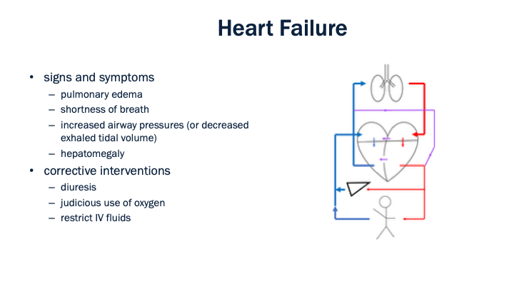
Another complication that we’ve previously noted is the development of heart failure. We first discussed this in the context of increased pulmonary blood flow and left-to-right shunts. At that time, we mentioned that this is something that needs to be monitored for, especially as the interventions we apply (i.e., withholding oxygen to increase pulmonary vascular resistance) can eventually contribute to the problem. We’ve outlines both signs and symptoms and also corrective interventions here.
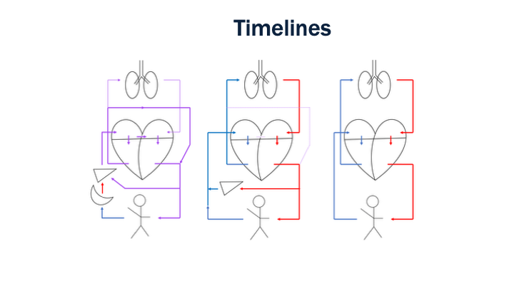
The final concept to discuss is the timeline at which we may come across these various defects. We already outlined the normal transition from fetal to neonatal to infant circulation, as pictured above, for the healthy child, but the timeline at which the transitions occur helps up identify which problems we might be up against for the child with a congenital heart condition. We don’t need to remember the specific components of the timeline, but we can generalize some trends.
Severe mixing lesions that might be incompatible with life typically present at the time of birth. Ductal dependent lesions with associated shock and/ or cyanosis often appear within the first month of life as collateral circulation (i.e. compensatory shunts) close off to blood flow. Other conditions, such as left-to-right shunts with atrial or ventricular septal wall defects present on the order of months after birth. In these patients, heart failure often develops gradually over time.
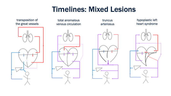
All that said, if we wanted to simplify all of this by timeline, here’s what we have to work with:
Cyanotic mixed lesions present soon after birth and our strategy is focused on maintaining oxygenation while preventing closure of the patent ductus arteriosus. We don’t always see marked evidence of increased or decreased pulmonary blood flow states, so our strategy if focused on keeping oxygenation in balance to both maintain delivery to the tissues and prevent closer of the PDA. And then is there is evidence of other things like heart failure, pulmonary edema, breath trapping, etc. we can address those as we already know how to do.
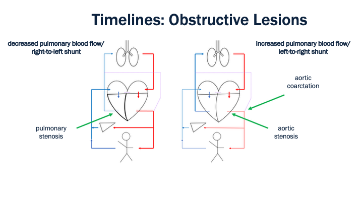
Obstructive lesions, on the other hand, have variable presentation depending on severity. Extreme cases will be evident in the first few days of life, while more subtle cases will not present until later in life – maybe not even until childhood or early adulthood. Treatment here is divided into our two contrasting strategies:
In those cases with decreased pulmonary blood flow and a right-to-left shunt we want to utilize interventions to increase pulmonary vascular resistance to ensure oxygenation and we monitor for heart failure. On the other hand, with increased pulmonary blood flow and left-to-right shunt we focus on decreasing pulmonary vascular resistance and ensuring that blood is delivered beyond the obstruction into systemic circulation.
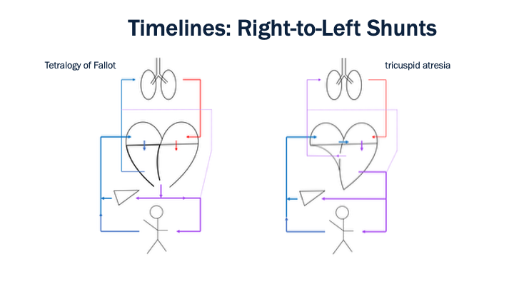
The right-to-left shunts will often present within the first month of life and are characterized by decreased pulmonary blood flow. Interventions focus on decreasing pulmonary vascular resistance and include oxygen administration, limiting PEEP and an increased minute volume with a lower-than-normal EtCO2 goal.
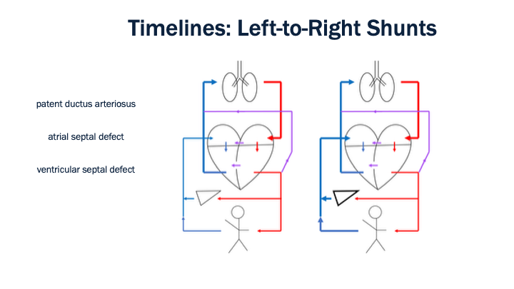
And lastly our left-to-right shunts typically present on the order of months. While the presentation of these defects is often associated with the onset of heart failure, interventions to address a diagnosed state of increased pulmonary blood flow focus on increased that pulmonary vascular resistance. Those interventions include limiting oxygen, utilizing PEEP, and decreasing minute volume with an EtCO2 higher than normal.
In the case that heart failure is already evidence, the strategy is then tailored to prevent hemodynamic consequences and includes diuresis and restricting IV fluids.
And again, it’s important to recognize that this discussion of timelines is variable depending on the severity of any given defect, these are simply generalizations we can use to help identify situations in an order to effectively apply interventions.
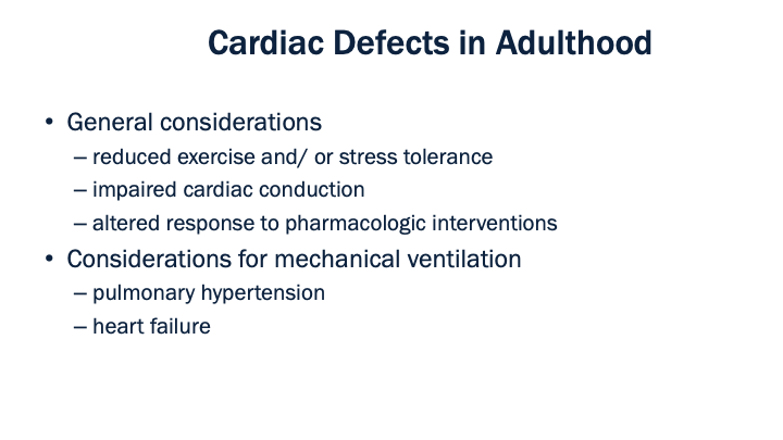
The very last thing to discuss is what to do if we have congenital heart defects that persist on through childhood and into adulthood. For the most part, congenital heart defects that are life threatening will be surgically treated in infancy and/ or childhood, so the acute nature of these conditions diminishes with time. It’s also the case that the more minor defects will resolve with time. That said, there are some things that may be present in the adult patient who needs mechanical ventilation and knowing which things may be encounters allows up to fine-tine our vent management strategies. It’s also the case that there are now more adults with congenital heart defects than in years past due to improved treatment strategies in childhood.
In general, adults with history of or newly diagnosed heart defects don’t require drastic changes in terms of mechanical ventilation strategies. It is worth noting, however, that there is an increased risk for both pulmonary hypertension and heart failure in this population. Interventions to address pulmonary hypertension are focused on decreasing pulmonary vascular resistance, just as with the neonate or infant – oxygen, avoid PEEP, aim low with minute volume. And as for heart failure, consider diuretics, restrict IV fluids, and be judicious with oxygen administration.